Supporting materials
Download
Download this article as a PDF


Microscope in Action is a hands-on educational resource for teaching fluorescence microscopy in the classroom and beyond
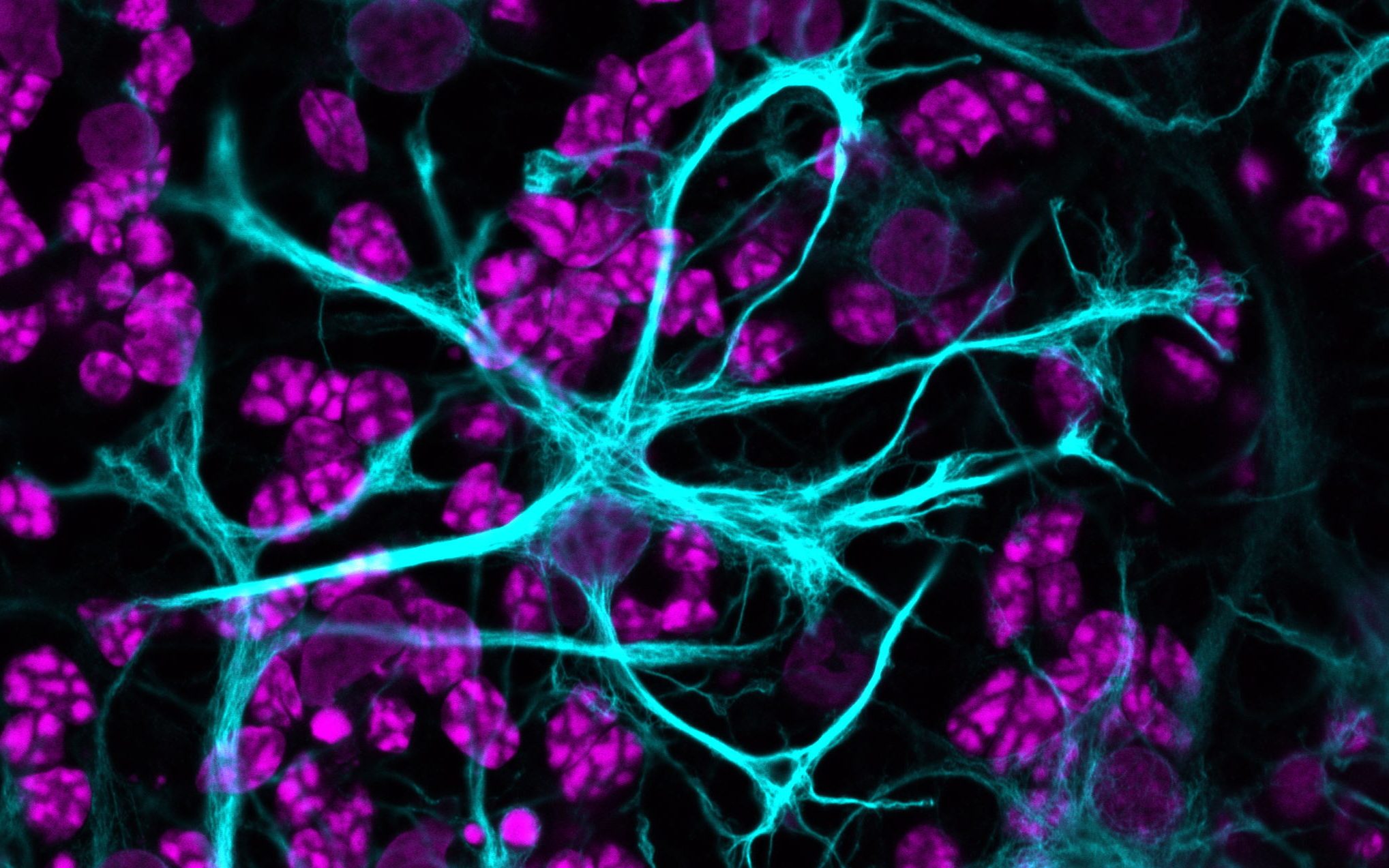
The development of fluorescence microscopy in the early 20th century marked the beginning of a new era in the field of microscopy and opened the door to a plethora of new discoveries. It enabled scientists to study the intracellular distribution of molecules like specific proteins and their interactions by using different fluorescent dyes and tags. It also made it possible to identify specific cell types in tissues and probe cellular dynamics, such as cell division, in living cells and organisms.
As a result, the fluorescence microscope is an indispensable tool for biologists. Despite its prevalence in life-science research, fluorescence is a neglected topic in secondary-school curricula. Furthermore, most fluorescence microscopes on the market are inaccessible to many schools. The European Learning Laboratory for the Life Sciences (ELLS), the education team of the European Molecular Biology Laboratory (EMBL), in collaboration with researchers from EMBL and EMBLEM Technology Transfer GmbH, decided to address this problem and developed the Fluorescence Learning Microscope as part of the comprehensive educational resource, Microscope in Action (MiA).
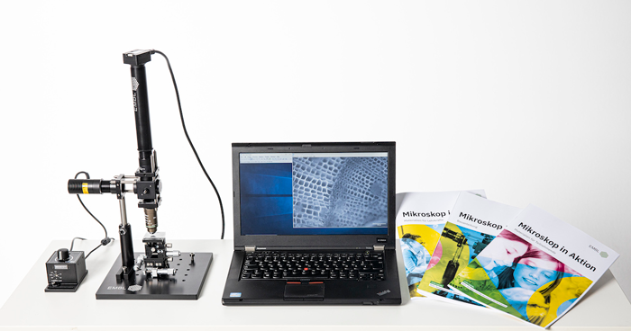
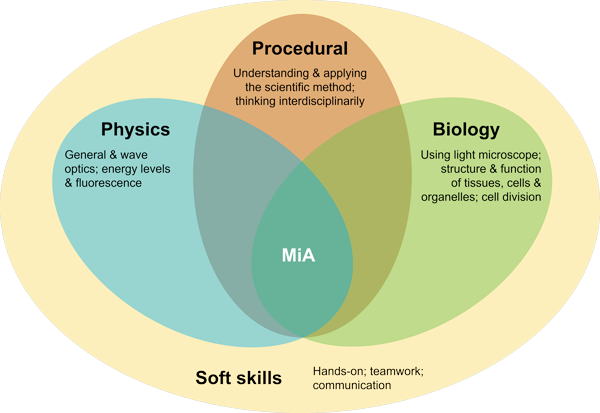
MiA is an educational resource targeted at secondary-school students. It contains a DIY fluorescence microscope called the Fluorescence Learning Microscope. Based on commercially available components, it is one of the most affordable research-grade fluorescence microscopes available. By employing pedagogical tools such as hands-on learning and collaborative problem solving, students can develop content knowledge in biology and physics while also increasing their competencies in procedural knowledge, such as applying the scientific method and thinking in an interdisciplinary manner. Students also improve key soft skills including teamwork and communication skills. Between 2018 and 2020, this newly developed resource was pilot tested in a series of classroom workshops.
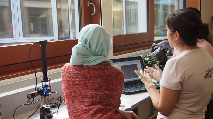
The MiA workshops are presented as three key modules: the assembly module, the sample preparation module, and the imaging module.
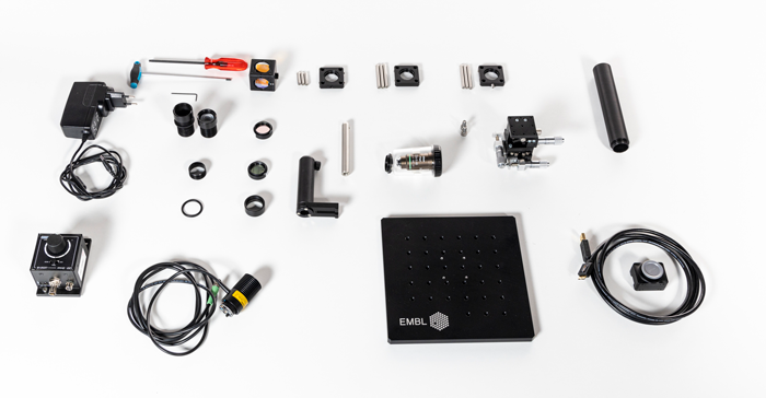
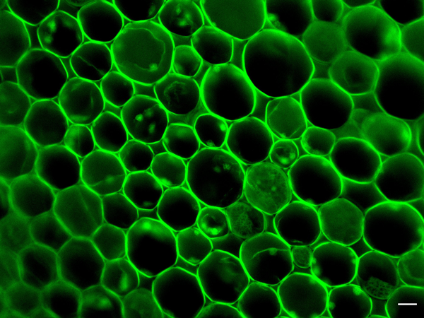
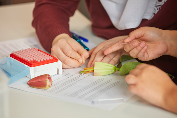
In the assembly module, the students put the fluorescence microscope together. The process of assembling the microscope helps them gain a deeper understanding of each individual component and its function. Once the microscope is assembled, the sample preparation module trains the students to prepare biological samples for imaging. This module includes various exercises which cover the basics of using a micropipette, preparing dilutions, mounting different materials like pollen or carrot sections on slides for observation, and the concept of staining.
MiA is designed to be flexible; teachers can use the various resources in whichever way fits their individual learning goals and caters best to the needs of their students. The handbooks include ready-to-use manuals, worksheets for classroom use, answer keys, extra assignments, and further reading sections. Additionally, teachers can utilize a ready-to-use slide deck to familiarize both themselves and their students with the basic concepts of fluorescence microscopy. Video guides provide support for microscope assembly and sample preparation and recorded talks by EMBL scientists can be used to introduce the topic in class and give insights into authentic life-science research.
To help teachers organise a MiA workshop in their classrooms, sample schedules are provided which range from one-day workshops to multi-lesson plans. In addition, the handbooks also offer recommendations on how to facilitate these sessions in groups to ensure that all students get the chance to partake in each and every aspect of the learning experience, from assembly to imaging.
Assessments and feedback of the learning experience showed that students displayed a marked improvement in practical and experimental skills and increased their learning motivation. Written in-class exams further indicated a strong grasp of the topics covered in the MiA sessions.
MiA is a versatile educational resource with curriculum links to physics and biology and connections to chemistry, technology, and art. It can be used in formal and informal learning environments with students from the age of 14. We offer a free-of-charge lease service through which you can bring the MiA experience to your classroom. This service is currently available to educators in Germany and is expected to expand to other countries soon. Further information about the resource and the lease service can be found on our webpage, https://www.embl.org/ells/microscope-in-action/.
If you would like to find out more about the science behind fluorescence microscopy, stay tuned for the upcoming Understand article brought to you by EMBL.
[1] Paci G. et al (2021) Microscope in Action: An Interdisciplinary Fluorescence Microscopy Hands-on Resource for Schools. The Biophysicist 2 : 55–73. doi: 10.35459/tbp.2020.000171
[2] Microscope in Action: https://www.embl.org/ells/microscope-in-action/
Articles on optics, fluorescence microscopy, and GFP
Articles on applications of fluorescence microscopy
Videos
«How inscrutable and incomprehensible are the hidden works of Nature!»
Antonie van Leeuwenhoek (1632 – 1723)
When Antonie van Leeuwenhoek perfected the lens used in seventeenth century to examine fabrics and used it to described red blood cells, spermatozoa, protozoa, and the bacteria found in stagnant water, biology was pushed into a new era, expanding the mechanistic view of the living world to the microscopic level. The discovery of microscopic beings conjured worlds of an infinitesimal scale… amazing and even bizarre worlds. Today, these worlds are not inscrutable and incomprehensible any more.
Microscope in Action, an affordable educational resource based on fluorescence microscopy, is a splendid idea and project, with the potential to propel the cell biology teaching at secondary school to a new level, both practically and conceptually. It offers an insightful, real-time approach to the core concepts of life. I hope that this initiative be rapidly and widely adopted in the European educational community.
Luis M. Aires, Biology teacher and education researcher, School Group Antonio Gedeao, Portugal
Download this article as a PDF