


Investigating the action of urease Teach article
Anna Lorenc from the Volvox project explains the importance of the enzyme urease and presents a protocol to demonstrate urease activity in the classroom.
Enzymes play an essential role in the metabolism of all organisms. They catalyse and control most biochemical reactions in our body – from the replication of genetic information (DNA polymerase) to digestion. Enzyme activity, however, is not always easy to visualise. This is a simple and cheap practical protocol to help teach the topic of enzyme activity in the classroom. All of the required materials are readily available and safe.
In this investigation, the enzyme urease from soya beans (Glycine max) breaks down urea to ammonia and carbon dioxide:
CO(NH2)2 + H2O → 2 NH3 + CO2
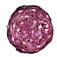
Ammonia (NH3) solution has a high pH which can be detected using a simple pH indicator, such as that obtained from red cabbage. The ammonia produced by the reaction can also be detected by smell.
As urease is produced by a wide range of different organisms, this practical activity can be used in lessons on:
- The nitrogen cycle
- The influence of organisms on their environment
- The adaptation of animals to different diets.
Background to the protocol
What is urea?
Every organism decomposes nucleic acids and proteins, generating nitrogenous waste because nucleic acids and proteins contain nitrogen. Mammals, amphibians and some invertebrates excrete nitrogenous waste as urea, which is produced in the liver. Urea is an especially good compound for disposing of nitrogen because it is water-soluble and less toxic than ammonia – the excretory produce of fish, for example. Human urine contains 2% urea.
Urea was also the first organic compound ever synthesised. In 1828, Friedrich Wöhler synthesised urea from inorganic compounds (lead cyanate and ammonium hydroxide). This was a landmark achievement: until then only living organisms were believed to be able to produce organic compounds, and these compounds were thought to be special and to require a ‘vital force’ to make them. Wöhler bridged the gap between the living and non-living worlds. He didn’t receive a Nobel Prize for his discovery though, because the Nobel Prize did not exist at that time. Today, urea is synthesised in vast quantities: it is used to make plastics and as a cheap nitrogenous fertiliser.
What is urease?
Urease catalyses the hydrolysis of urea to carbon dioxide and ammonia. It is found mainly in seeds, micro-organisms and invertebrates. In plants, urease is a hexamer – it consists of six identical chains – and is located in the cytoplasm. In bacteria, it consists of either two or three different subunits. For activation, urease needs to bind two nickel ions per subunit.
How did urease become famous?
Urease from jack beans (Canavalia ensiformis) was the first enzyme ever purified and crystallised, an achievement of James B. Sumner in 1926, at a time when most scientists believed that it was impossible to crystallise enzymes. This earned Sumner the 1946 Nobel Prize in Chemistry. Today, crystallisation of proteins helps scientists to discover their structure and determine how they work. This knowledge permits the design of substances that interfere with enzyme action, such as the anti-AIDS drugs which inhibit the action of HIV’s enzymes or recent developments towards a possible rabies treatment (Ainsworth, 2008).
Why is there urease in soya beans?
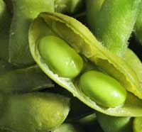
Image courtesy of iStockphoto
The role of urease in soya beans is not entirely clear, although it is possible to speculate. Soya leaves also contain urease, but here, the enzyme is a thousand times less active than in the beans. It is known that the leaf enzyme helps to recycle nitrogen from proteins (the proteins are broken down to urea). In the beans, urease does the same when the beans germinate. The resulting ammonia from the reaction may also protect the plant cells from pathogens – it seems that the plant enzyme itself is an insecticide.
Where else can urease be found?
Many species of bacteria produce urease, including Helicobacter pylori, the bacterium responsible for stomach ulcers. By doing this, H. pylori raises the pH of the gastric juice from about pH 3 to pH 7, the optimal pH for its growth. A commercially available test for H. pylori checks for the presence of urease in breath, and is used as a tool for diagnosing stomach ulcers.
Ruminants (such as cows and sheep) have cellulose-digesting bacteria in their rumen – the first compartment of their stomachs – to help them digest their plant diet. Ruminants excrete urea into this part of the stomach, the urea making an excellent source of nitrogen for bacterial growth. To take up the nitrogen, the bacteria secrete urease to break down the urea. Eventually, the animals digest the bacterial mass.
Is urease in soya beans harmful to humans?
Urease is not harmful. However, raw soya beans contain other compounds which are unhealthy. For example, there is a protein inhibitor in raw soya beans which prevents the digestive enzyme trypsin from working and makes raw soya beans inedible. The presence of the inhibitor is not easy to detect, but fortunately, it has a similar level of heat intolerance to urease – both are inactivated by heating. Therefore, to ensure that the inhibitor is inactivated, commercial preparations of soya beans (soya flour or foods that contain soya, such as tempe and tofu) are tested for urease activity – in a very similar way to the test described here. If no urease activity can be detected, the inhibitor has presumably also been inactivated.
Urease in the nitrogen cycle
Nitrogen is a crucial element for plant growth, but most plants can only use it in the form of ammonium or nitrate. Only legumes (thanks to the bacteria they live in symbiosis with) and cyanobacteria can use elemental nitrogen from the air.
Many animals excrete urea in their urine. Soil micro-organisms feed on animal urine, producing urease to transform the urea to ammonia, which is then readily accessible to plants. This is part of the nitrogen cycle, the process by which the nitrogen from proteins and other compounds is constantly recycled.
The protocol
This protocol allows students to detect the activity of a plant enzyme in seeds. When the substrate (urea) is present, urease breaks it down into carbon dioxide and ammonia. Dissolved in water, ammonia raises pH, an effect seen with the red cabbage pH indicator. In the experiment, students observe that to obtain the product (ammonia), both a substrate (urea) and an enzyme (urease) are needed. They observe that the enzyme activity raises the pH.
Red cabbage extract – a great pH indicator
In this protocol, we use red cabbage extract as a pH indicator. It contains anthocyanins. The structure and colour (when in solution) of these compounds are pH-sensitive. At pH 7, the solution is violet/blue; in the acidic range it turns red. When the pH rises above 7 and the solution is more alkaline, the extract turns green. These colour changes are reversible – just check what happens when you add citric acid and baking soda (sodium bicarbonate) one after another.
Materials and equipment
For each student or group of students:
- 20 ml 10% solution of fertiliser urea (a solid fertiliser made of pure urea)
- 5 ml 10% solution of citric acid (or other low-pH solution)
- 5 ml 10% solution of sodium bicarbonate (NaHCO3) (or other high-pH solution)
- 40 ml red cabbage indicator, prepared as described below
- 10 ml soya bean extract, prepared as described below (from 1 g of dried soya beans)
- 10 ml distilled water
- Pasteur pipettes or a plastic drinking straw, for transferring the solutions
- 6 test tubes and a test tube rack.
For preparing soya extract and red cabbage indicator, a blender and boiling water are needed, as well as coffee filter paper and a funnel.
Timing
The experiment can be completed in 30 minutes.
The soya bean suspension and red cabbage extract can be made in advance, or their preparation can be demonstrated during the lesson. This takes only ten minutes, but note that the soya beans must be soaked in water for at least one hour before the lesson.
The reaction between soya urease and urea also takes about ten minutes.
Preparation
Red cabbage indicator
- Cut two large red cabbage leaves into strips and place them in a heat-resistant container such as a beaker. Pour on 200 ml of freshly boiled water, soaking the leaves completely.
- After 5 min, decant the liquid and leave it to cool.
- Throw the leaves away.
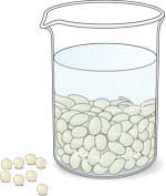
Soya bean extract containing urease
- Soak the soya beans in water for at least 1 h (preferably overnight).
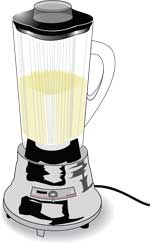
- Blend the soaked soya beans in a food blender with about 10 ml of water per gram of dry soya (the water from soaking the beans can be used) until the mixture is smooth (for about 5 min).
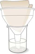
- Filter the soya purée through filter paper in a funnel.
- Collect and keep the filtrate, which contains urease.
Investigation
The volumes given below are for standard (~10 ml) glass test tubes.
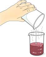
First, ask the students to check how pH influences the colour of the red cabbage extract.
- Pour 3 ml of red cabbage indicator into each of three tubes.
- Add 2 drops of citric acid solution to the first test tube. Mix and observe the colour change. Rinse the pipette with water after the addition of each component.
- Add 2 drops of sodium bicarbonate solution to the second test tube. Mix and observe the colour change.
- Add 2 drops of distilled water to the third test tube. Mix and observe the colour change.
- Compare and note the colours of the solutions in all three test tubes. Put the tubes aside to act as a reference.
Next, investigate the effect of urease from soya beans on urea.
- Add 2 ml of red cabbage indicator to each of three fresh tubes.
- Mix 2 ml of urea solution each into two of these tubes. Note the colour.
- Add 2 ml of the soya suspension each to the remaining tube and to one of the tubes with urea solution.
- Compare the colours and odours of the mixtures in all tubes. Repeat this observation after 3 min.
Interpretation
Red cabbage indicator turns green with increasing pH. This colour change, along with an ammonia odour, can be detected in a tube containing both the enzyme and its substrate. In tubes containing either enzyme only or substrate only, the pH stays stable, so the red cabbage indicator stays violet.
Safety
Approximately 1% of children may be allergic to soya bean extract (see McGee, 2004). Teachers are advised to check that none of their students is affected before attempting this protocol.
Further investigations
A follow-up experiment could be conducted to investigate factors influencing enzyme activity (such as temperature, pH and the concentrations of enzyme, substrate and product).
Acknowledgements
This practical protocol was developed by Anna Lorenc for the Volvox project, which is funded by the Sixth Framework Programme of the European Commission.
The Volvox project
The Volvox project team is a group of biology teachers and specialists from ten countries across the European Union, aiming to provide secondary-school biology teachers and others with proven laboratory protocols, simulations, classroom activities and numerous other educational resources. See www.eurovolvox.org
References
- Ainsworth C (2008) Locking the cradle. Science in School 8: 25-28. www.scienceinschool.org/2008/issue8/rabies
- McGee H (2004) On food and cooking. London, UK: Hodder & Stoughton. ISBN: 0340831499
Review
Enzyme activity is frequently investigated using the action of amylase on starch, which links to food and nutrition. The simple method presented here makes a good change that links to the nitrogen cycle, which can be a difficult concept for students to grasp. The idea of using a natural indicator that students can isolate themselves makes the lesson more fun. Also, students can smell the product of the reaction due to the release of ammonia! Factors affecting enzyme activity could be investigated as group work and this could be discussed in a plenary session.
The article could be used to test comprehension by asking questions such as:
- Why are the cabbage leaves placed in boiling water?
- What colour will the indicator turn if ammonia is present in the solution?
- Describe what you would see if you used boiled soya bean extract and explain why this happens.
- State which of the reaction tubes is the ‘control’ experiment. Explain what this means and what you would expect to observe in this tube.
Shelley Goodman, UK