Bringing the beauty of proteins to the classroom: the PDB Art Project Teach article
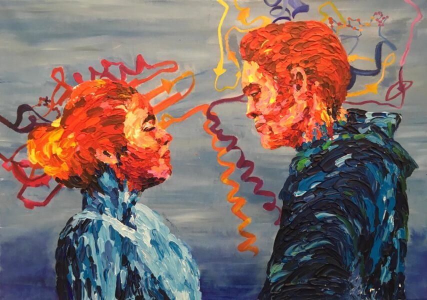
The PDB Art project aims to make science more accessible and inspire young people to explore the beauty of proteins by bringing together art and science.
© PDBe/EMBL-EBI
The Protein Data Bank in Europe (PDBe) celebrated its fifth edition of the PDB Art project in December 2020. This initiative brings together art societies, school children, arts and science departments, scientists, and members of the PDBe team. This collaborative effort inspires school students to create artworks based on proteins – life’s building blocks – while introducing them to the world of structural biology. Not only do students create truly outstanding pieces of art, but they also provide a wider perspective of the impact of science upon society through their thoughtful interpretation of the chosen protein or topic of interest.
We highly recommend that art and science departments work together collaboratively, as this tends to give more successful project outcomes. Although this method requires greater strategic planning, the involvement of both science and art teachers helps to provide expertise across both subject areas and leverage the interdisciplinary nature of the project.
An example of this collaborative approach was adopted by teachers from Thomas Gainsborough School in Suffolk, UK, where the art and science departments worked together to develop materials for art and science lessons. The Thomas Gainsborough School have kindly allowed us to share these materials, which are available from the PDB Art Resources page.
If a collaboration is not possible, schools can run the project within individual subject lessons, and the PDBe team can provide scientific support. Alternatively, it is possible to involve local scientific staff, for example, through STEM hubs, or local equivalents, and local university networks.
How is the PDB Art project structured?
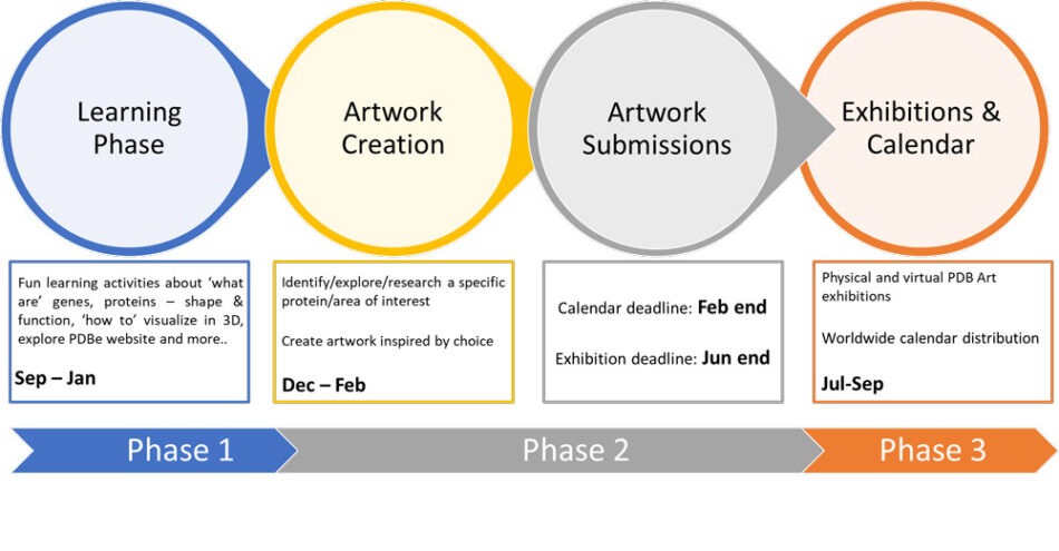
The PDB Art project timelines are aligned with the academic school year; however, there is a large amount of flexibility to support involvement in the project. Participating pupils come from a range of different year groups, from year 7 (11–12 years old) to year 12 (16–17 years old), allowing the project to be run in a classroom setting or as a more independent research project. Choosing the year group depends on the school and is at the teacher’s discretion. The PDB Art project has three phases: learning phase, creation and submission phase, and celebration phase. Help and support from the PDBe is provided throughout, if needed. The project timelines and details are shown below.
Phase 1 – Learning and idea development
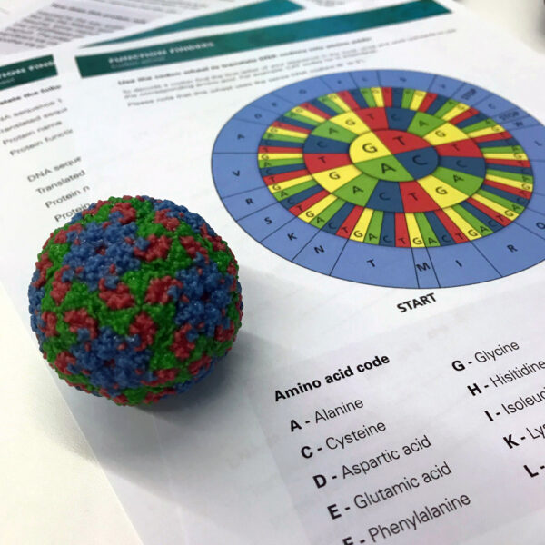
Image courtesy of Ben Keeble/The Perse School/Cambridge/UK
The PDB Art project starts with the learning phase, where students learn, explore, and gain an understanding of protein structure and function, which can be achieved by using PDBe activities or following the normal school curriculum and supplementing it with the PDBe resources listed below. All these activities and resources have been put together by PDBe scientists to support learning and build interest and confidence in understanding protein structures.
Activity 1 – Function finders
Through this activity, students will learn about the concept of genes and how they encode proteins.
Materials
- Printed handouts for the function finders activity
- YourGenome translation/transcription video, as an initial introduction
- The provided PowerPoint presentation for teachers
Procedure
- The PowerPoint presentation provides an outline for teacher on how to run the activity and gives details of each of the proteins found in the Protein Profiles booklet.
- Students are provided with their own DNA sequence and codon wheel printed sheets. Using the codon wheel, students translate a DNA sequence into the representative protein sequence. This helps them to understand the processes of transcription and translation.
- Students then use the Protein Profiles booklet to find ‘their’ protein and learn more about its function, for example, learn about bioluminescence by looking at the luciferase enzyme found in fireflies and other bioluminescent organisms.
- Finally, students use the PDBe structure link on the worksheet to access the 3D view of the protein structure on a phone, tablet, or computer.
Activity 2 – Exploring protein structure
Materials
- A set of videos prepared by PDBe to help students the importance of proteins
- 3D modelling kit (recommended)
Procedure
- Students should watch the videos to understand the importance of proteins in our everyday life.
- Using the amino acid starter kit, students are given a protein ‘toober’ and amino acid clips. They add amino acids randomly onto the toober. Following the properties of amino acids on the accompanying amino acid board, students fold up the protein toober into a 3D shape.
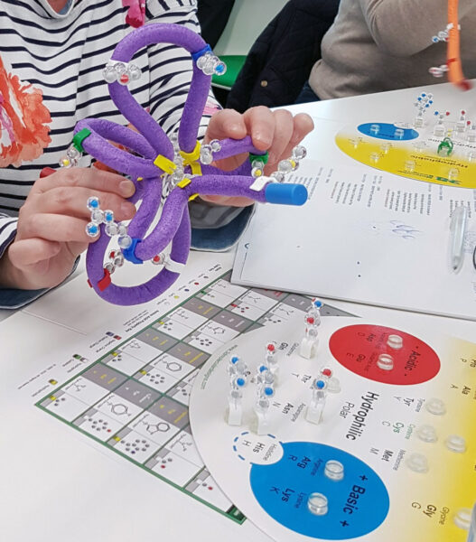
Image courtesy of Deepti Gupta
At PDBe, we have a selection of kits (Water Kit, Enzymes in Action Kit, Substrate Specificity Kit, Flow of Genetic Information Kit, Amino Acid Starter Kit and Protein-Folding Kit) available to lend out, although availability depends on demand.
Activity 3 – Using the PDBe website and choosing a protein
In textbooks, proteins are often shown as 2D images, and while this can be a good way to understand the basic principles, proteins truly come alive when seen in 3D. Seeing how they are arranged and interact in 3D gives a fascinating insight into the tiny world of these biological molecules, like the spike protein from the SARS-CoV-2 virus shown below.
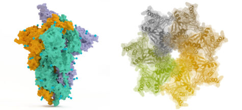
Image courtesy of Deepti Gupta and David Armstrong
Materials
- Introductory worksheet
- Pinterest activity worksheet
- Computers/tablets
Procedure
- Students should work through the introductory worksheet, which guides students through using the PDBe website. This will allow them to explore and visualize biomolecules in 3D and learn about their functions.
- They should then use the PDBe website to search for known proteins, such as insulin or haemoglobin.
- Students should then explore different proteins and choose a ‘protein of interest’ for their art project. The hard part is narrowing down their options and choosing just one of the interesting structures to investigate from the thousands available!
- Students can take a look at our featured structure articles for some more ideas. They can even travel back in time and view structures originally determined back in the 1970s.
- To help students decide which proteins to focus on, the PDBe has created a dedicated PDB Art Pinterest account, providing ideas for specific proteins and scientific topics. Students should use the Pinterest activity worksheet to guide them through finding proteins of interest and learning how to view the structure in 3D using the PDBe website. Students learn to visualize proteins in 3D and see them in different forms.
Phase 2 – Creating art from protein structures
From the activities above, students make an informed choice for their protein of interest and move on to the art project, where they create an artwork with support from their teachers.
The choice of the type of artwork has no prescription. This flexibility gives students the freedom to visualize and explore new forms of art and be more innovative in their representations. It also provides a more accessible and unique approach to learning the scientific concepts. As Albert Einstein once said, “The greatest scientists are artists as well. Arts and sciences are branches of the same tree”.
Paintings, drawings, etchings, digital art, photograms, sculptures, textile pieces, ceramics, batik designs, and even a musical piece have been created so far. All these can be explored on the PDB Art webpage.
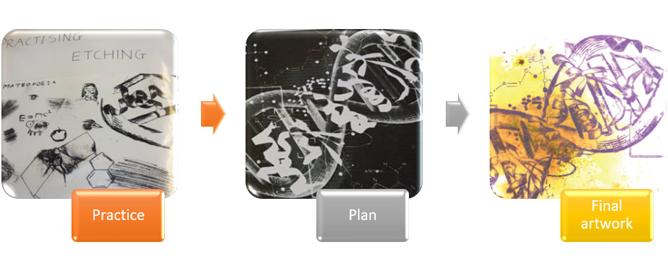
Image courtesy of Deepti Gupta and David Armstrong
Materials
- Art supplies
- Computers/tablets for research and viewing of 3D structures
Procedure
- Students should read about the function of their chosen protein, explore available structures at the PDBe, and understand its importance in biology and society.
- Students should then generate ideas for an artwork and start working on their piece.
- Towards the end of this process, they should write accompanying descriptions for their artwork, explaining which protein they were inspired by, why they found it interesting/important, how they represented it, and what media they chose.
- Students may also provide video interviews explaining the background of their artwork and what inspired them to create the piece.
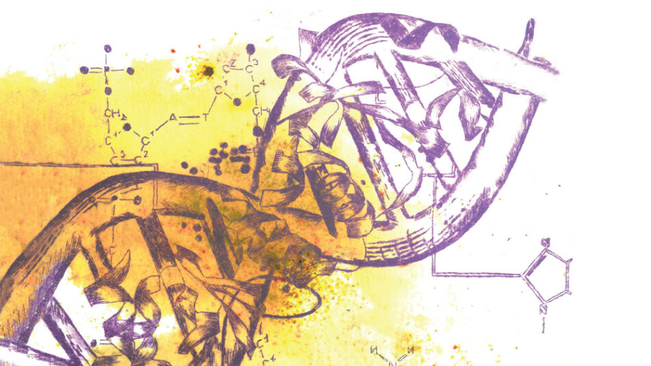
A high-resolution digital copy of the artwork and description should be submitted to the PDBe by the end of February for consideration for the yearly PDBe calendar. All artwork created by students should be submitted electronically by the end of June for consideration for the PDB Art exhibition.
Phase 3 – Celebrating the artworks
One of the key stages of the project is sharing the artworks with the public and with scientists around the world, through exhibitions and other methods. The PDBe hosts public art exhibitions, both physically and virtually, to showcase and celebrate these remarkable creations. The exhibition’s private opening event provides a platform to spark discussion and engagement among students, scientists, artists, and the public.
In addition, the annual PDBe calendar, featuring the artworks, is distributed worldwide, bringing artistic interpretation back to the scientific community. In 2021, the PDB is celebrating 50 years of archiving structures of biological molecules with an anniversary edition of the calendar.
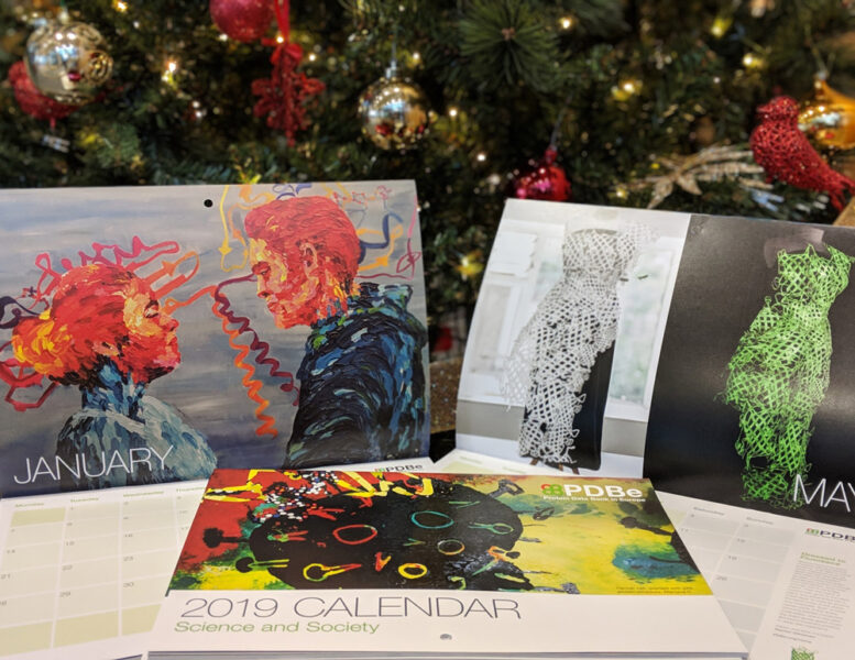
Image courtesy of Deepti Gupta and David Armstrong
PDBe calendars and artworks exhibited from previous years can be explored on the PDB Art webpage.
How to get involved
The PDBe can help you get started with the PDB Art project and provide you with support in running activities at your own school.
If reading this article has inspired you to join the PDB Art project, then fill in the ‘Expression of Interest’ form or send an email to pdb-art@ebi.ac.uk.
We can then discuss the project further with you and introduce you to our community of teachers already running the project.
Acknowledgements
The cover picture for Issue 54 is based on the artwork by Lucy Kerr, Y12 student of Thomas Gainsborough School. The PDB id used for the artwork is 5F1S.
Resources
- Find out more about the PDB Art project.
- Discover more teaching materials on the PDB Art Resources page.
- Watch student testimonials and webinars on the PDBe YouTube channel.
- Read an introductory article on the PDB art project: Gupta D, Armstrong D (2021) Introducing students to the beauty of biomolecules. Science in School53.
- Discover how artificial intelligence is helping to predict protein folding: Heber S (2021) From gaming to cutting-edge biology: AI and the protein folding problem. Science in School52.
- Find out how art can be inspired by molecular biology: Stroe O (2019) Art meets molecular biology. Science in School46:29–33.
- How can we see proteins in action? Wilson R (2021) Plant solar power: unlocking the secrets of photosynthesis with X-ray free-electron lasers. Science in School54.
- Read about the background of structures highlighted on the PDB page.
Institutions
Review
Excellent ideas for interdisciplinary projects – learning about the importance of proteins and using the knowledge to inspire art creations.
The article could be used to stimulate discussion about molecular biology and the importance of proteins as building blocks.
Marie Walsh, Science Lecturer, Ireland


